(submitted 30 June 2022; revised 02 July 2022; accepted 03 July 2022)
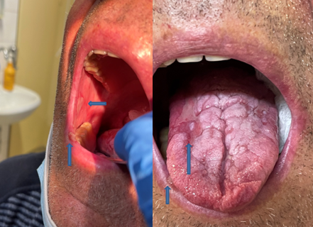
Image. Careful examination of the oral cavity discovered findings indicative primarily of a systemic disease. Our patient’s, oral mucosa and tongue (blue arrows) had features of discoid lesions, characterized by a well-demarcated zone of erythema, silvery white, scarred plaques atrophy, ulceration accompanied by radiating striae. These lesions resemble those found in patients with erosive lichen planus. Also a discoid lesion was noted on the inferior lip vermilion and in the periodontal gingival mucosa and bilaterally on the buccal. Although many organs can be affected in patients with of systemic lupus erythematosus (SLE), cutaneous lesions are seen in the great majority. In our case, early oral manifestation was not accompanied with skin or other visceral manifestations. Serologic identification of anti-double stranded DNA (ds-DNA) and anti-Sm antibodies, the serological autoantibody markers of SLE, have assissted in the firm diagnosis of the autoimmune rheumatic disease.
AUTHORS CONTRIBUTION
The author prepared the manuscript and the artwork. The author approves the final version of the manuscript.
Τα cookies είναι σημαντικά για την εύρυθμη λειτουργία του ιστοτόπου και την βελτίωση της online εμπειρίας σας. Πατήστε "Αποδοχή" για να συνεχίσετε!ΑποδοχήΠερισσότερες πληροφορίες