A 40-year-old lady presented in the Department of Rheumatology and Clinical Immunology of our Hospital with low grade fever over a two months period and tenderness over the sternum, initially thought to be costochondritis by a general practitioner.
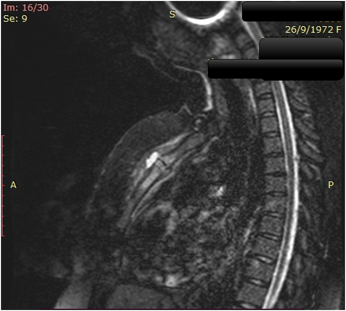
Contrast enhanced sagittal T2-weighted magnetic resonance imaging (MRI) of chest showed high signal intensity of bone marrow at the angle of Louis (sternal angle) the upper part of the body of the sternum and the lower half of the manubrium. Inflammatory tissue with enhancement was also seen at anterior – presternal region.
While bacteriological culture results were pending, antibiotic therapy with Staphylococcus aureus coverage were initiated empirically for treating primary sternal osteomyelitis. The aetiologic agent was Staphylococcus hominis and no apparent risk factor was detected. Primary sternal osteomyelitis is a rare clinical entity.
Fig. 1 Contrast enhanced sagittal T2-weighted magnetic resonance imaging (MRI) of chest showed high signal intensity of bone marrow at the angle of Louis (sternal angle) the upper part of the body of the sternum and the lower half of the manubrium (red arrow).
AUTHOR CONTRIBUTION
The author prepared the manuscript and the artwork. The author approves the final version of the manuscript.
Τα cookies είναι σημαντικά για την εύρυθμη λειτουργία του ιστοτόπου και την βελτίωση της online εμπειρίας σας. Πατήστε "Αποδοχή" για να συνεχίσετε!ΑποδοχήΠερισσότερες πληροφορίες